


 |
| 3D pictures of All Stages of Mitosis cell division |
MITOSIS:
For eukaryotic cells in general, the life cycle extends from the time of the cell`s formation until its own subsequent division is completed.Mitosis is a very small part of this cycle; it only lasts for few minutes or an a hour or more.Normally, the non dividing cell spends about ninty percent of its life in Interphase, when its increase it mass, approximately double the number of its cytoplasmic components, and finally duplicates its DNA. Interphase actually consists of three phases of activities: the gaps were so named before we knew much about what was going during the cell cycle.
 |
| Initial preparation stages of Mitosis |
We know that cells must have built-in instructions regarding the duration of a cell cycle, because the duration of cell cycle, because the duration is fairly consistent for all the members of a species. Sometimes adverse environmental conditions do arrest cells in G1 phase.(For example, this happens among ameobas and other Protistans when they are deprived of vital nutrient.) Even so, if a cell progresses beyond a certain point in G1 (The so called restriction point), the cycle normally will be completed regardless of outside conditions.
 |
| Chromosome Number in Mitosis |
 |
| Microscopic pictures of stages of Mitosis cell division |
DIFFERENT PHASES OF MITOSIS
 |
| 3D Picture of Interphase |
INTERPHASE:
Two daughter nuclei are formed, each with a diploid number of chromosomes (The same as the parental nucleus).
 |
| 3D Picture of Early Prophase |
EARLY PROPHASE:
The DNA and associated proteins start condensing into the threadlike chromosome form. (Chromosomes are already duplicated.) Two chromosomes derived from themale parent are in blue ; their homologues from the female are red.
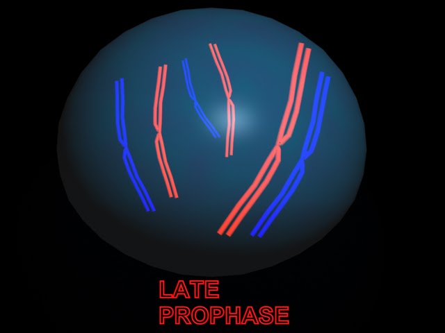 |
| 3D Picture of late prophase |
LATE PROPHASE:
Chromosomes continue to condense.Microtubule start to assemble outside the nucleus; they will form the spindle,which will establish poles for division.The nuclear envelope starts breaking up centrioles(if present) are moved by the microtubules toward opposite poles.
 |
| 3D Picture of Metaphase |
METAPHASE:
Nuclear envelope is gone; spindle penetrates region of nucleus . Sister chromatids of each chromosome are attached at their kintochores t the spindle. All the chromosomes are lined up at the sindle equator.
 |
| 3D Picture of Anaphase |
ANAPHASE:
Sister chromatids of each chromosome will now be separated from each other and moved to opposite poles.
 |
| 3D picture of Telophase |
TELOPHASE:
Chromosomes decondense.New nuclear membranes start forming.Most often, cytokinesis occurs before the end of telophase.
3D Animation of Mitosis Cell Division created by Me (Manash Kundu)
 |
| 3D pictures of All Stages of Meiosis cell division |
MEIOSIS:
It took billions of cell division to produce all the tissues of your body, and every day millions of cells still divide every second just to replace worn-out or damaged predecessors and to keep that body running smoothly. Most are somatic cells; they are the ones making up all body tissues expect for the germ cells, a cell lineage set aside for sexual reproduction.
sexual reproduction begins with meiosis and proceeds through the formation of gametes ( sex cells, such as sperms and eggs). It ends at fertilization, when the sperm nucleus and egg nucleus fuse in the first cell of the new individual (the zygote). A zygote grows into a multi celled adult by way of mitosis, which faithfully maintains the chromosome number characteristic of the species, division after division For example, there are 46 chromosomes in your somatic cells, 48 in Gorillas, and 14 in garden peas with each mitotic cell division, that number does not change.
 |
| Number of Chromosomes during Meiosis |
Meiosis only occurs in germ cells that are destined a give rise to gametes. With this division mechanism, the chromosome number is not maintained; it is reduced in half. and not just any half - each gamates ends up with of each pair of homologous chromosomes. ( It does not matter which of the two it gets.) It takes Two nuclear divisions to put one chromosome of each type into each gamete: Thus gamates end up with a haploid number of chromosomes, meaning "half" the parental number. Then when a sperm nucleus and an egg nucleus fuse at fertilization, the diploid number is restored. " Diploid" means having two chromosomes of each type (that is, pairs of homologous chromosomes) in the somatic cells of sexually reproducing species.
DIFFERENT PHASES OF MEIOSIS:
 |
| 3D picture of Prophase-1 |
PROPHASE 1:
Each chromosome condenses, then pairs with its homologue crossing over and recombination occurs.
 |
| 3D picture of Metaphase-1 |
METAPHASE 1:
Spindle apparatus has formed and nuclear envelops broken down during transition to metaphase 1. Homologues align randomly at spindle equator; some spindle microtubules attach each pair of homologues to opposite poles.
 |
| 3D picture of Anaphase-1 |
ANAPHASE-1:
Each hmlogue is separated from its partner, and the two are moved to opposite poles.
 |
| 3D picture of Telophase-1 |
TELOPHASE 1:
A haploid number of chromosomes (still duplicated) ends up at each pole.
 |
| 3D picture of Prophase-2 |
PROPHASE-2:
There are no DNA replication interkinesis, which precedes prophase 2 in most species.Sister chromatids of each chromosome are still attached at the centromere during this often brief stage.
 |
| 3D picture of Metaphase-2 |
METAPHASE 2:
Each chromosome is aligned at the spindle equator; microtubules attach its sister chromatids to opposite poles.
 |
| 3D picture of Anaphase-2 |
ANAPHASE 2:
Each chromosome splits what were once sister chromatids are now chromosomes in their own right and are moved to opposite poles.
 |
| 3D picture of Telophase-2 |
TELOPHASE 2:
Four daughter nuclei form; each has a haploid number of chromosomes, all of which are in the unduplicated state.
3D Animation of Meiosis Cell Division created by Me (Manash Kundu)
Microtubules:
These are larger contractile protein fibres that are involved in movement of
Organelles within the cell
Chromosomes during cell division
Cell extensions
Centrosome:
This directs organisation of microtubules within the cell.It consists of a pair of centrioles (Small clusters of microtubules)
and plays an important role during cell division.
 |
| Microscopic pictures of stages of Mitosis cell division |
 Nature Magazine
 National Geographic
 Cartoon Games
 History Channel
 Online Movies
 Scientific American
 Filmfare Mag
 Online Britannica
 Time Mag
 Unexplained Mysteries


No comments:
Post a Comment