 | |
| Animated Lymphatic System |
 |
| Human Lymphatic System With all Organs |
LYMPHATIC SYSTEM:
All body tissue are bathed in tissue fluid,consisting of the diffusible constituents of blood and waste materials from cells.Some tissue fluid returns to the capillaries at their venous end and the remainder diffuses through the more permeable walls of the lymph capillaries,forming lymph. Lymph passes through vessels of increasing size and a varying number of lymph nodes before returning to the blood.The lymphatic system consists of:
 |
| Human Lymphatic System with all its Described parts |
Lymph.
Lymph vessels.
lymph nodes.
Lymph organs e.g. spleen and thymus.
diffuse lymphoid tissue, e.g. tonsils.
Bone marrow.
Functions of lymphatic system:
Tissue drainage:
Every day, around 21 liters of fluid from plasma carrying dissolved substances and some plasma protein, escape from the arterial end of the capillaries and into the tissues.Most of this fluid is returned directly to the bloodstream via the capillary at its venous end, but 3-4 lit of fluid are drained away by the lymphatic vessels. Without this the tissue is rapidly become waterlogged, and the cardiovascular system would begin to fail as the blood volume falls.
Absorption in the small intestine:
Fat and the fat soluble materials. e.g. the fat soluble vitamins, are absorbed into the central lacteals (lymphatic vessels) of the villi.
Immunity:
The lymphatic organs sre concerned with the production and maturation of lymphocytes the white blood cells responsible for immunity .The bone marrow is therefore considered to be lymphatic tissue,since lymphocyte are produced there.
LARGER LYMPH VESSELS:
The walls of lymph vessels are about the same thickness as those of small veins and have the same layer of tissue,e.g. the fibrous covering, a middle layer of smooth muscle and elastic tissue and an inner lining of endothelium .Lymph vessels have numerous cup-shaped valve to ensure that lymph flows in the one way only i.e. toward the thorax.There is no pump like the heart, involved in the onward movement of lymph but the muscle layer in the walls of large lymph vessels has an intrinsic ability to contract rhythmically(The lymphatic pump).
In addition, any structure that periodically compresses the lymphatic vessels can assist in the movement of the lymph along the vessels,commonly including the contraction of adjacent muscles and the pusation of large arteries.
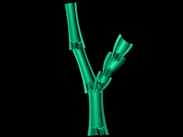
Lymph vessels become larger as they join together,eventually forming two large ducts, the thoracic duct and right lymphatic duct, which empty lymph into the subclavian veins.
Thoracic duct:
This duct begins at the cysterna chyli, which is a dilated lymph channel situated in front of the bodies of the first two lumbar vertebrae.The duct is about 40 cm long and opens into the left subclavian vein in the root of the neck.It drains lymph from both legs, the pelvic and abdominal cavities, the left half of the thorax, head and neck and the left arm.
Right lymphatic duct:
This is a dilated lymph vessel about 1 cm long.It lies in the root of the neck and opens into the right subclavian vein. It drains lymph from the right half of the thorax, head and neck and the right arm.
 |
| Lymph Nodes with all its Described Parts |
LYMPH NODES:
Lymph nodes are oval or bean-shaped organs that lie, often in groups, along the length of lymph vessels.The lymph drains through a number of nodes, ususally 8 to 10, before returning to the venous circulation.These nodes vary considerably in size: some are as small as a pin head and the largest are about the size of an almond.
 |
| Another Picture of Lymph Node |
Structure of lymph nodes:
Lymph node have outer capsule of fibrous tissue that dips down into the node substance forming partitions or trabeculae.The main substance of the node consists of reticular and lymphatic tissue containing many lymphocytes and macrophages.
As many as 4 or 5 afferent vessel carries lymph away from the node.Each node has a concave surface called the hillum where an artery enters and a vein and the efferent lymph vessel leave.
The large number of lymph nodes situated in strategic positions throughout the body are arranged in deep and superficial group.
Lymph from the head and neck passes through deep and superficial cervical nodes. Lymph from the upper limbs passes through nodes situated in the elbow region,then through the deep and superficial Axillary nodes.
Functions of lymph nodes:
1)Filtering and phagocytosis:
Lymph is filtered by the reticular and lymphoid tissue as it passes through lymph nodes.Particulate matter may include microbes, dead and live phagocytes containing ingested microbes, cells from malignant tumours, worn out and damage tissue cells and inhaled particles. Organic material is destroyed in lymph nodes by macrophages and antibodies. Some inorganic inhaled particles cannot be destroyed by phagocytosis.These remain inside the macrophages,either causing no damage or killing the cell.Material not filtered out and dealt with in one lymph node passes on to successive nodes and by the time lymph enters the blood it has usually been cleared of foreign matter and cell debris.
2) Proliferation of lymphocytes:
Activated B and T -lymphocytes multiply in lymph nodes. Antibodies produced by sensitised B-lymphocytes enter lymph and blood draining the node.
 |
| Spleen with all its described parts |
SPLEEN:
The spleen contains reticular and lymphatic tissue and is the largest lymph organ. The spleen lies in the left hypochondriac region of the abdominal cavity between the fundus of the stomach and the diaphragm.It is purpish in colour and varies in size in different individuals,but is usually about 12 cm long, 7cm wide and 2.5 cm thick.It weight about 200 g.
Structure:
The spleen is slightly oval in shape with the hilum on the lower medial border.The anterior surface is covered with peritoneum.It is enclosed in a fibroelastic capsule that dips into the organ,forming trabeculae.The cellular material,consisting of lymphocytes and macrophages,is called splenic pulp, and lies between the trabeculae.Red pulp is the part suffused with blood and white pulp consists of areas of lymphatic tissue where there are sleeves of lymphocytes and macrophages around blood vessels. The structure entering and leaving the spleen at the hilum are:
Splenic artery, a branch of the coeliac artery.
Splenic vein, a branch of the portal artery.
Lymph vessels(efferent only).
Nerves.
Blood passing through the spleen flows in sinuses,which have distinct pores between the endothelial cells, allowing it to come into close association with splenic pulp.
 |
| Lymph Vessels |
Functions:
Phagocytosis:
As described previously , old and abnormal erythrocytes are destroyed in the spleen,and the breakdown products bilirubin and iron are transported to the liver via the splenic and portal veins. Other cellular material, e.g. leukocytes,platelets and microbes is phagocytosed in the spleen.Unlike lymph nodes, the spleen has no afferent lymphatics entering it, so it is not exposed to diseases spread by lymph.
Storage of Blood:
The spleen contains up to 350 ml of blood, and in response to sympathetic stimulation can rapidly return most of thisvolume to the circulation,e.g. in haemorrhage.
Immune response:
The spleen contains T- and B-lymphocytes, which are activated by the presence of antigens,e.g. in infection.Lymphocyte proliferation during serious infection can cause enlargement of the spleen (splenomegaly).
Erythropoiesis:
The spleen and liver are important sites of the fetal blood cell production, and the spleen acn also fulfil this function in adult in times of great need.
 |
| Thymus Gland with all its Described Parts |
THYMUS GLAND:
The thymus consists of two lobes joined by areolar tissue the lobes are enclosed by a fibrous capsule which dips into their substance,dividing them into lobules that consist of an irregular branching framework of epithelial cells and lymphocytes.
Function:
Lymphocytes originate from pluripotent stem cells in red bone marrow.Those that enter the thymus develop into activated T-lymphocytes.Thymic processing produces mature T-lymphocytes that can distinguished self tissue from foreign tissue,and also provide each T-lymphocyte with the ability to react to only one specific antigen from the millions it will encounter.T-lymphocytes then leave the thymus and enter the blood. Some enter lymphoid tissues and other circulate in the bloodstream.T-lymphocyte production,although most prolific in youth,probably continues throughout life from a resident population of thymic stem cells.The maturation of the thymus and other lymphoid tissue is stimulated by thymosin, a hormone secreted by the epithelial cells that form the framework of the thymus gland.Involution of the gland begins in adolescence and with increasing age,the effectiveness of the T-lymphocyte response to antigens declines.
All The 3D Pictures in This Post are created by Me (Manash Kundu)


 Online Movies
Nice blog, keep sharing such posts. Best breast disease treatment center provides integrated and individually customized solutions for treating lymphatic disorders. Book your one-on-one consultation today.
ReplyDeletelymphatic massage San Diego
This comment has been removed by the author.
ReplyDeleteVery informative and helpful post. Best surgery care treatment center provides integrated and individually customized solutions for treating lymphatic disorders. Book your one-on-one consultation today.
ReplyDeleteFat Disorders San Diego